Object Description
3D Print of a heart with Atrial Septal Defect was printed in 2017 by the Department of Radiology, University of Ottawa / Medical Imaging, The Ottawa Hospital Dr. Frank Rybicki, Dr. Adnan Sheikh and Dr. Leonid Chepelev printed this model, as well as several other 3D models of organs for the Touch section of the Medical Sensations Exhibit at the Canada Science and Technology Museum.
It is based on this case study.
The object, a blue triangular and detailed item, is quite bright, especially contrasting with its lightwood and white background.
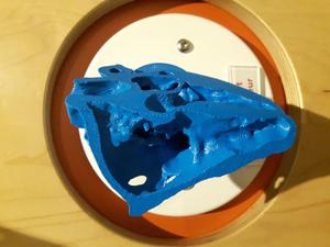
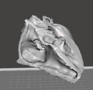
When I first saw the object, I could not automatically determine what is was, especially from the angle I had approached. Upon further observations, I can detail the object’s design as bumpy, full of holes and crevices, rigid surfaces on the outside, with softer curved ripples on the inside. The inside of the object is quite unique. Because of its curvaceous design and the (probably) way it was created, this creates unique lines inside the object, resembling thumbprints.
Both visually and when examined through touch, the object is not symmetric. It's texture and complexity make it a challenging object for study using 3D software, and printing.
For the exhibit, Dr. Leo Chepelev designed the 3D print with a cylinder base of 1½ inch height and a 3-inch diameter. This cylinder supports the object by attaching itself to the back of the object, in its rounded out back.

Photo: Ingenium: Canada's Museums of Science and Innovation The origins of this 3D print in Dr. Leonid Chepelev's own words:
"Basically, this was a congenital case with multiple abnormalities, and we used the model to design a patch for a hole inside the heart, which would be just one of several procedures that this child would undergo. The model is meant to demonstrate what's called an Atrial Septal Defect, or in other words, a hole between two of the four chambers of the human heart, the left and the right atria. The heart surgeons close these defects (holes) using a patch of material which should be of appropriate size, and this model helps them do just that. Because congenital heart malformations are often very unique, it is difficult to get a large number of cases for heart surgeons to become very experienced with every type of malformation. So, models like the one that we are showcasing really help the surgeons to practice the complex procedures they will perform before actually opening up the patient. Keep in mind - when the patient is on the operating table in heart surgery, the heart is stopped and there is a large oxygen exchange/pump machine that takes over the circulation. The less time is spent during a procedure such as this, the better, so these models really help the surgeons and the patient.
To make a model like this, we give the patient some contrast to make their heart and vessels bright, and we scan the patient using a CT scanner. We then take those pictures which basically give us 1mm slices of the patient over the part that we are looking at, and we use them to make a model just like described in the paper above."
Exerpt from Email to David Pantalony, Feb 10, 2018
